Simplified View of Fascia
Fascia fills the spaces in the body so that there can be movement along different anatomical structures and fluid exchange between them. These spaces spread throughout the body connecting different body components to form what has been recognized as another body organ, the interstitium.
It is often pointed out that the white layer under the skin of raw chicken is an example of fascial tissue. Its structure consists of two collagen layers that connects to other structures with a viscous web between these layers that stretches and changes with movement. There are various components that exist in the fluid that fills this space along with nerves and blood vessels. The properties of this fluid varies in response to numerous factors. This in turn affects the quality (stiffness) of the fascia.
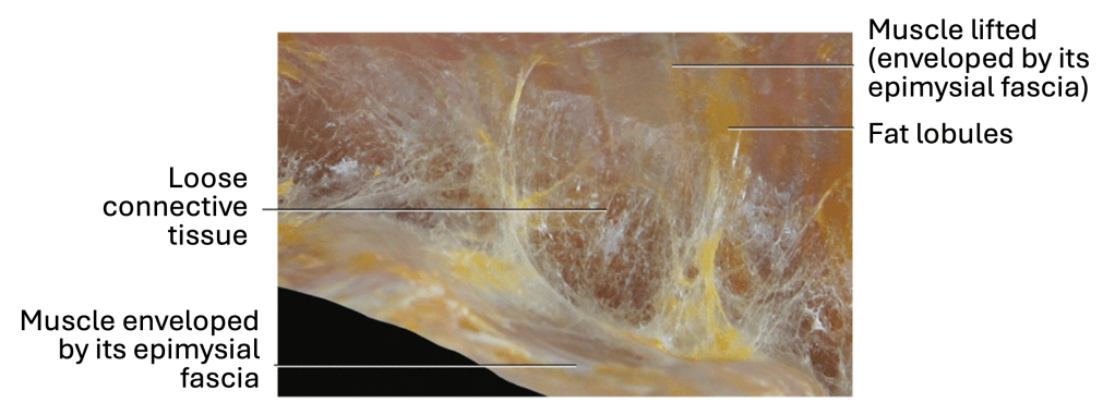
Figure 1: Macroscopic aspect of the loose fascia between pectoralis major and minor muscles. The loose fascia creates a gliding surface between the two muscles and permits their independent contraction.
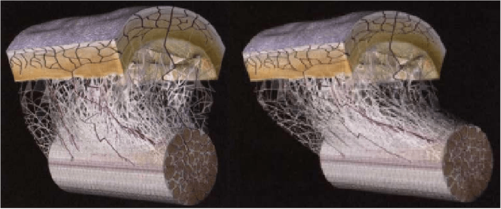
Figure 2: Computer generated images of the fascial membrane between a tendon and its surrounding carpal sheath based on microscopic videos with the impact of sliding shown in right hand side
Fascia Classification of Interest
One way fascia can be classified is the location of the tissues.
Types of fascia of current interest include:
•Superficial Fascia – Between skin and fatty tissue below skin.
•Intermuscular Septum – Separates certain muscles and compartments of muscles.
•Epimysium – wraps individual muscles.
•Perimysium – wraps bundles of muscle fibers within the muscle.
•Endomysium – wraps individual muscles fibers.
•Periosteum – wraps the bones.
Not Shown in figure:
•Tendons – Extension of the various muscle fascia as they attach to the bone.
•Ligaments – Could be consider extensions of the periosteum between bones.
•Visceral Fascia – Surrounds the internal organs
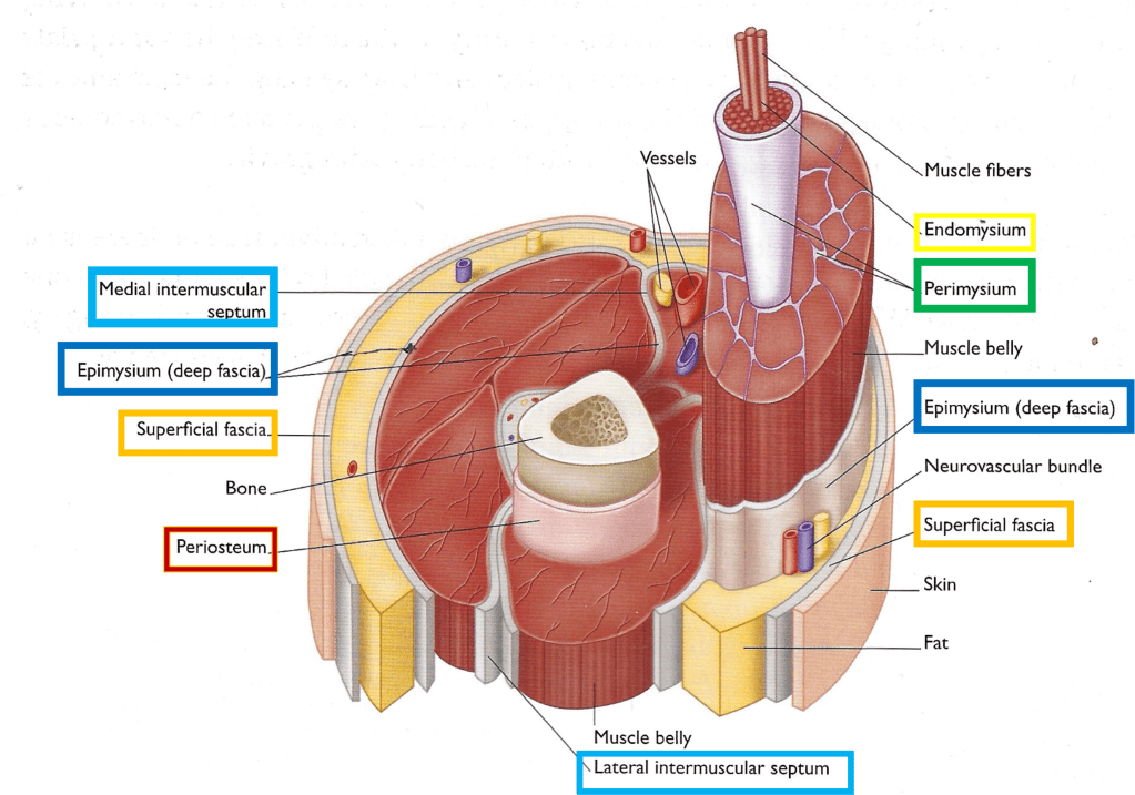
Figure 3: Various types of fascia are highlighted with colored boxes to provide some perspective about the pervasiveness of fascial tissue. This cross section example uses raised components to help identify anatomy related to fascial system
Movement & Fascia
Fascia has many incredible properties including the ability to respond to both local, distal, and global changes. It helps immobilize joints in response to injuries. With training, it enables the performance of elite athletic. It provides a degree of hydraulic pressure for fluid movement through the body. It contain ions that carry charges. There is evidence the Chinese median system utilizes pathways within the fascia.
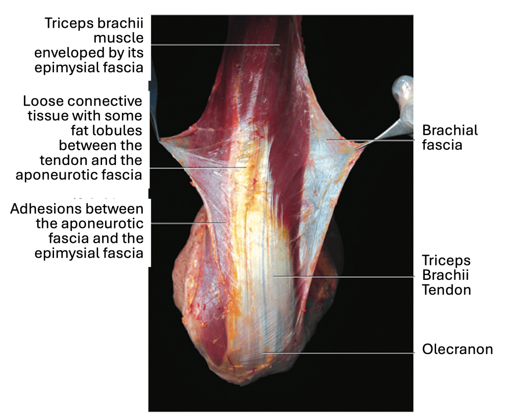
Figure 4: The brachial fascia is an aponeurotic fascia that allows freer movement of the muscles and their epimysial fascia below.
Index: Science of Tai Chi Poster Info
References:
Figure 1: Stecco, C. (2014). Functional Atlas of the Human Fascial System .
Figure 2: Guimberteau, J. C. (2012). The subcutaneous and epitendinous tissue behavior of multimicrovacuolar sliding system. In R. Schleip, T. W. Findley, L. Chaitow, & P. Huijing (Eds.), Fascia: The Tensional Network of the Human Body(pp. 143-146). Edinburgh: Churchill Livingstone Elsevier.
Figure 3: Earls, J. (2014). Born to Walk. Chichester, England: Lotus Publishing.
Figure 4: Stecco, C. (2014). Functional Atlas of the Human Fascial System .
© 2026 James F Sturnfield
