Insight between Tàijíquán & Fascia
Many Tai Chi teachers utilize fascia research to explain various aspects of the art. These explanations are sometimes in conflict with each other and tend to lean on single aspects of Tai Chi and fascia. Still, this shows the power of fascial research that helps bring a deeper understanding of Tai Chi. These understandings can help teachers train beginners for more consistent health benefits and to reduce injuries that can occur in Tai Chi training.
I am not aware of much utilization of Tàijíquán principles to advance fascia research. This can only begin to happen as advancements in biomechanical measurements provide deeper insights into how Tai Chi manipulates the body. This requires studies done with advanced practitioners of the art.
Various aspects of fascial research
Numerous views can be found of how Tàijíquán utilizes the body. Some of these have been mathematically modeled to different degrees. Others are currently more conceptual models.
Anatomy Train model
Anatomy trains, also known as myofascial meridians, are intended to describe the pathways of functional force that travels through the body. There are 12 sets of these anatomy trains identified in the different layers of the fascia.
This chain concept extends the muscle and bone model of movement to longer connections through the body with many additional insights and challenges.
Figure 1: Some lines of the anatomy train are shown here. The deep arm lines are shown on the left with front and back lines, the superficial arm lines are on the right
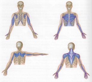
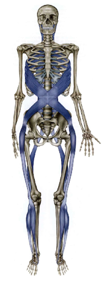
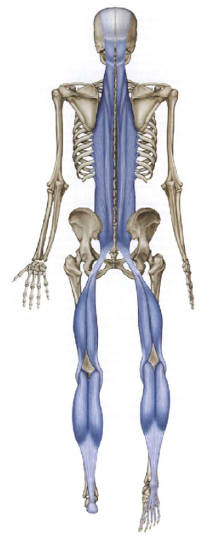
Figure 2: The front view of the spiral lines on the right while the back extension of these lines are shown on the left.
Biotensegrity model
The basic concept is for a healthy body, the bones float in a balance of tension created by the soft tissue. The detailed mechanism is still being developed. This probably needs to involve the structure of the joint capsule, the interactions of different regions of the fascia, and the muscle fiber.
It is a challenge to generate a model with the correct properties and correspondence to the current understanding of the human anatomy.
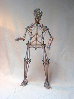
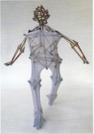
Figure 3: Biotensegrity model that approximates the human body by Tom Flemons
Hydrodynamic Model
The fluid of the fascia lies between two collagen layers that are connected by a ”webbing” of collagen fibers. The properties of this fluid varies, exerting different forces around the body influenced by numerous factors.
Including these factors into a model can provide a more realistic understanding of the interaction of Tai Chi and fascia.
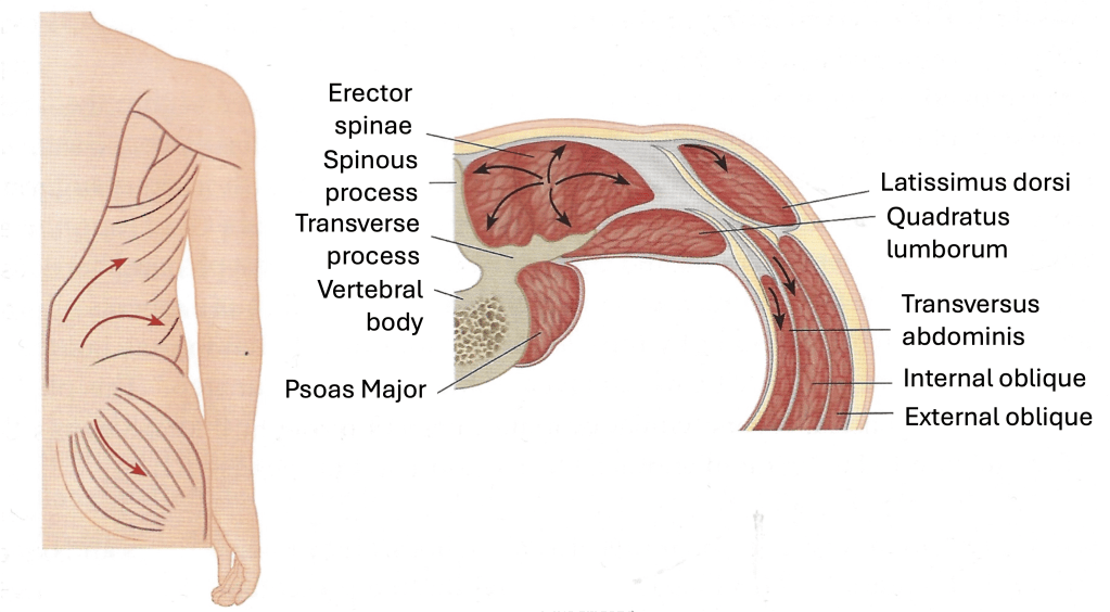
Figure 4: When the gluteus fascia and latissimus dorsi contract, tension is created in the surrounding fascia pushing them out, which in turn creates matching force in underlying muscle that supports the hips and knee.
Membrane Model
The fascia are membranes that intersect each others as they wrap around and fill the spaces in the body. These can be mathematically modeled using flexible body analysis and similar methods. The membranes can sometime modeled as 2-dimensional objects with varying properties to simplify calculations.
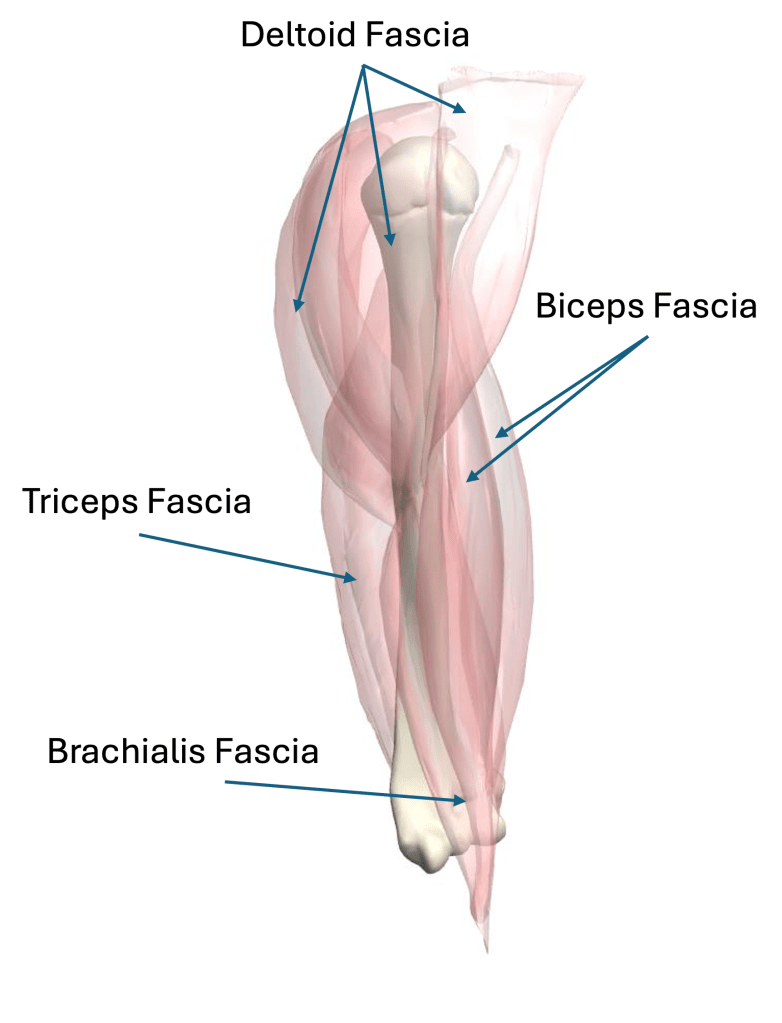
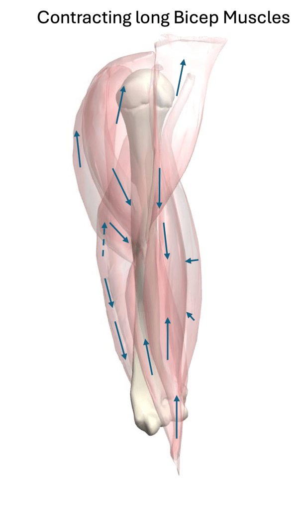
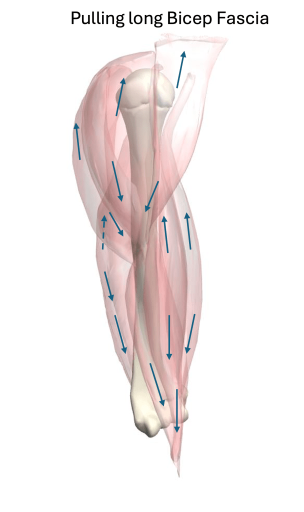
Figure 6: Some of the epimysium fascia of the upper arm and their response as a membrane due to a contraction of the long bicep muscle compared to a stretching of the muscle.
Balloon Model
The balloon model has several different variations of design and provides a simplified combination of a membrane with a hydrodynamic model. In most versions, the superficial fascia is treated as a balloon with varying elastic properties. In one case, the epimysium is shaped by contractions of muscle fibers. In another case, the epimysium is also treated as balloons. There is a viscous fluid that fill the space of the balloons.
Previous: Biomechanic View for Tàijíquán
Index: Science of Tai Chi Poster Info
References:
Figure 1: Lesondak, D. (2018). Fascia: What it is and why it matters. Pencaitland, East Lothian, Scotland: Handspring Publishing Limited
Figure 2: Myers, T. W. (2021). Anatomy Trains: Myofascial Meridians for Manual Therapists & Movement Professionals 4th Ed.Elsevier Limited.
Figure 3: Lesondak, D. (2018). Fascia: What it is and why it matters. Pencaitland, East Lothian, Scotland: Handspring Publishing Limited
Figure 4: Earls, J. (2014). Born to Walk. Chichester, England: Lotus Publishing.
Figure 5: Earls, J. (2014). Born to Walk. Chichester, England: Lotus Publishing.
© 2026 James F Sturnfield
